Posted by: Skin And Cancer Institute in Medical Dermatology
We’ve identified granuloma annulare as a chronic inflammatory dermatosis caused by delayed-type hypersensitivity reactions, often triggered by trauma, infections, or autoimmune conditions like diabetes. Treatment approaches include topical corticosteroids for localized lesions, intralesional injections every 6-8 weeks, and narrowband UV-B phototherapy for generalized forms. Advanced options involve JAK inhibitors like baricitinib, systemic therapies including methotrexate, and biological agents for refractory cases. Understanding the specific subtype and underlying triggers will optimize your therapeutic strategy.
Key Takeaways
- Granuloma annulare has unclear causes but involves immune responses triggered by trauma, infections, and autoimmune conditions.
- Topical corticosteroids and intralesional injections are first-line treatments for localized lesions, requiring repeated applications.
- Generalized cases respond to narrowband UV-B phototherapy and systemic medications like hydroxychloroquine or methotrexate.
- JAK inhibitors like baricitinib show rapid effectiveness within 1-2 months for treatment-resistant cases.
- Approximately 50% of localized cases resolve spontaneously within 24 months without any medical intervention.
What Is Granuloma Annulare and How Does It Present
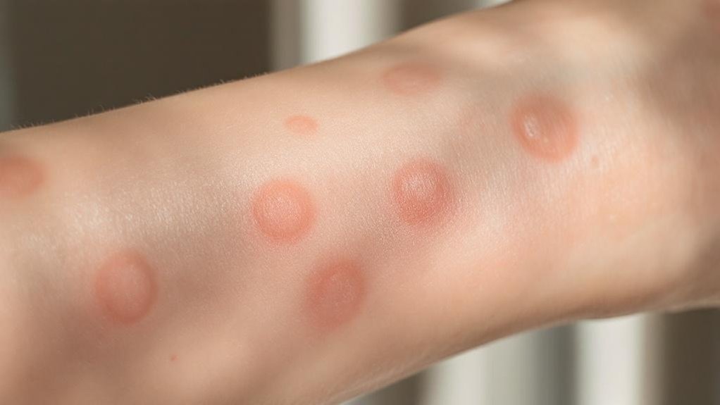
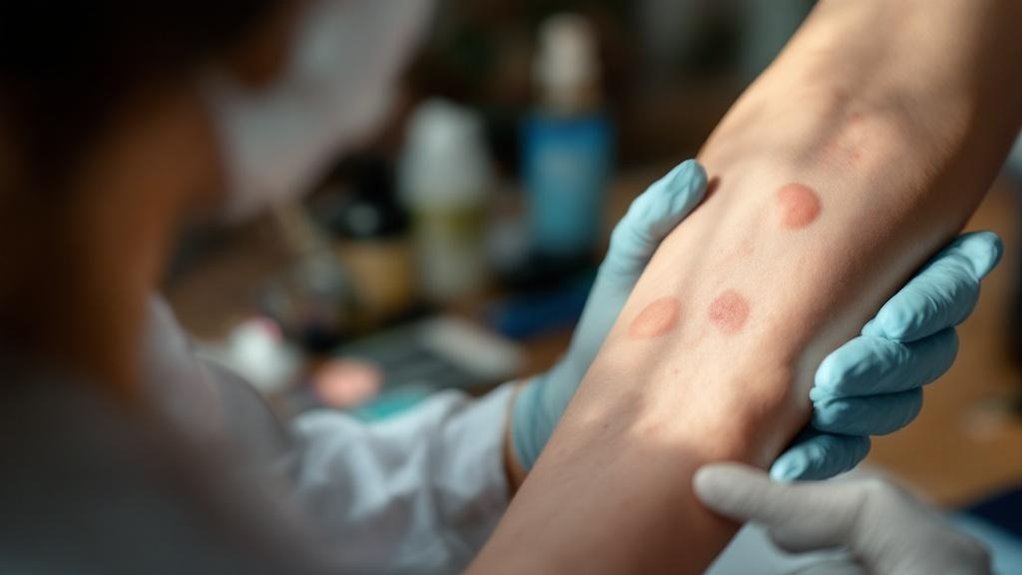
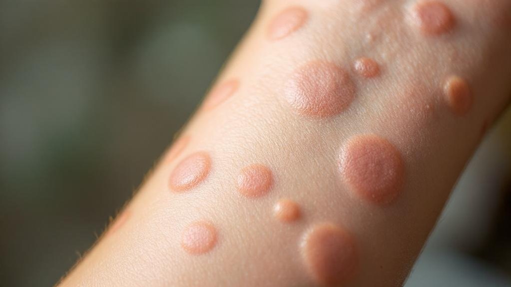
Granuloma annulare represents a chronic, benign inflammatory dermatosis characterized by distinctive ring-shaped arrangements of papules and plaques. We recognize that identifying this non-contagious condition can be challenging for patients experiencing its unique presentation. The granuloma characteristics include smooth, firm bumps measuring 1-2 mm in diameter that form circular patterns expanding to 2.5-5 cm. Lesion appearance varies considerably, ranging from reddish, pink, and violet to skin-colored or yellow presentations. We’ve observed that localized forms mainly affect the backs of hands and forearms, while generalized variants involve multiple body regions. Unlike many inflammatory conditions, these lesions typically don’t present with scaling. Most patients experience minimal symptoms beyond cosmetic concerns, though mild itching occasionally occurs. The condition shows a female predominance with slightly higher occurrence rates in women compared to men.
Understanding the Underlying Causes and Risk Factors
While the precise etiology of granuloma annulare remains elusive, we’ve identified it as a multifactorial inflammatory response pattern rather than a single disease entity. The causal mechanisms center on delayed-type hypersensitivity reactions mediated by Th1 immune responses and tumor necrosis factor alpha-driven inflammation. Environmental triggers include minor trauma, insect bites, sun exposure, and viral infections such as Epstein-Barr virus and HIV. We’ve documented significant associations with autoimmune conditions, particularly thyroid disease and type 1 diabetes. The 2:1 female predominance suggests hormonal influences, while HLA marker frequencies indicate genetic predisposition. Two main theories regarding its cause involve tissue breakdown by immune cells or an immune overreaction to harmless substances. Understanding these interconnected factors helps us appreciate why certain patients develop granuloma annulare while providing essential insights for targeted therapeutic approaches.
Different Types of Granuloma Annulare and Their Characteristics
Because granuloma annulare presents through distinct morphological patterns, we’ve classified this condition into five primary subtypes that differ considerably in clinical presentation, patient demographics, and diagnostic considerations. Localized granuloma annulare affects hands, feet, and wrists with circular lesions up to 5 centimeters. The generalized variant involves multiple body areas with generalized itchiness and poorly-defined rings exceeding 10 centimeters. Subcutaneous nodules characterize the subcutaneous subtype, primarily affecting children aged 2-5 years across various lesion locations including extremities and scalp. Patch type lesions appear as flat, discolored areas without distinct ring formations. Perforating granuloma annulare demonstrates perforating characteristics with central umbilication and potential crusting. Each subtype requires specific diagnosis confirmation methods, with excisional biopsy often necessary for subcutaneous presentations. The condition is notably more common in females than males across all subtypes.
Diagnostic Methods for Accurate Identification
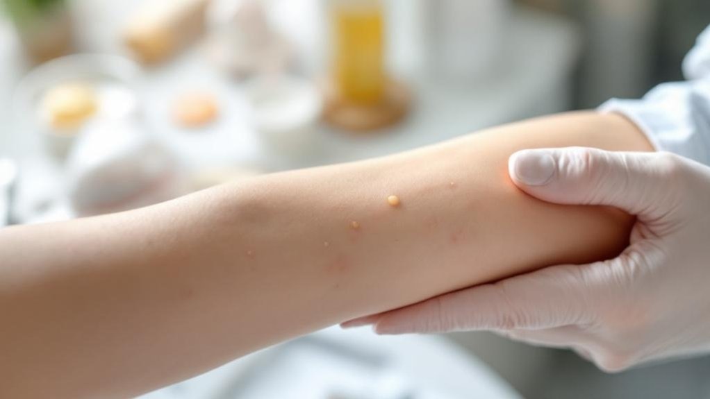
At the Skin and Cancer Institute, we employ systematic visual examination techniques to identify the characteristic annular ring of firm, flesh-colored papules that define granuloma annulare, documenting their distribution on extremities and noting the absence of epidermal scaling that differentiates this condition from other annular dermatoses. When clinical presentation suggests granuloma annulare, we proceed with biopsy confirmation to reveal the pathognomonic palisading granulomas with central collagen degeneration and mucin deposition visible through special stains like Alcian blue. Our thorough diagnostic approach combines dermoscopic evaluation to identify structureless white zones and vascular patterns with ancillary testing including skin scrapings to exclude fungal infections and laboratory investigations for associated systemic conditions. In most cases, our dermatologist diagnosis requires minimal testing due to the distinctive clinical appearance of the red rings with normal-appearing skin in the center.
Visual Examination Techniques
Accurate identification of granuloma annulare begins with systematic visual examination of the characteristic non-scaling plaques that typically present on the dorsal aspects of extremities. We assess the annular arrangement of firm, shiny papules that appear violaceous, erythematous, or flesh-colored with central depression. Our examination focuses on symmetrical presentation patterns and expanding bumps that join to form circular lesions. We differentiate from tinea corporis by confirming the absence of peripheral scaling, a key diagnostic feature. Visual documentation captures the lesion’s morphology for accurate diagnosis and patient education regarding prognosis. We evaluate the localized variant’s discrete ring-shaped formations while noting that subcutaneous variants manifest as firm lumps beneath the skin surface rather than raised surface rashes. The classic appearance includes a beady annular border with central delling, though macular presentations may lack raised components entirely.
Biopsy Confirmation Process
While visual examination provides valuable diagnostic information, definitive confirmation of granuloma annulare requires histopathological analysis through tissue biopsy. We utilize specific biopsy techniques based on clinical presentation—excisional biopsy serves as our standard approach for subcutaneous variants, while punch biopsy proves essential when dealing with clinically ambiguous scenarios.
Our histopathological analysis reveals characteristic diagnostic markers: palisading epithelioid histiocytes surrounding necrobiotic collagen, focal collagen degeneration, and mucin deposition within dermal layers. We’ve documented four distinct patterns—interstitial (57.9%), palisaded granulomatous (26.3%), sarcoidal granulomatous, and mixed variants. The presence of anuclear dermis further supports the diagnostic confirmation in typical cases.
Diagnosis confirmation necessitates correlating clinical findings with histologic features. For disseminated forms, we may need multiple biopsy sites to guarantee accurate identification and differentiation from similar conditions like necrobiosis lipoidica.
First-Line Treatment Approaches and Medications
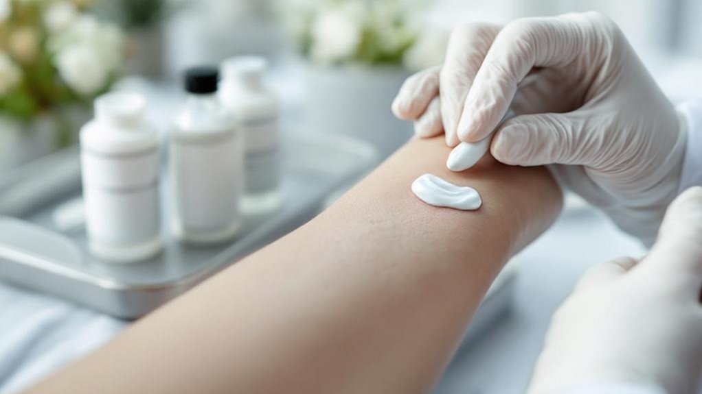
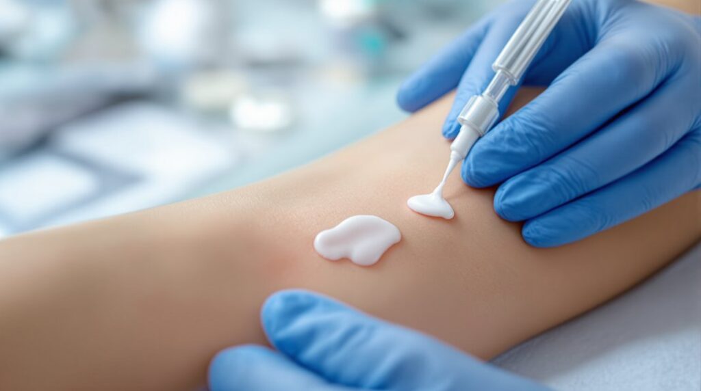
Although granuloma annulare affects only 0.06% of the US population, we’ve established several effective first-line treatment approaches that target both localized and generalized variants of this condition. For localized lesions, we recommend topical corticosteroids as standard therapy, with prescription-strength formulations accelerating clearance when combined with occlusive dressings. Intralesional injections represent our most widely recognized first-line intervention, requiring repeat treatments every 6-8 weeks until resolution occurs.
When managing generalized cases affecting extensive body surface areas, we utilize narrowband ultraviolet B phototherapy for its favorable long-term risk profile. Antimalarial medications like hydroxychloroquine serve as our primary oral first-line option for widespread disease. While no FDA-approved therapies exist specifically for granuloma annulare, these evidence-based approaches provide reliable outcomes for our patients. The JAK-STAT pathway involvement in granuloma annulare pathogenesis has opened new avenues for targeted therapeutic interventions.
Advanced Therapeutic Options for Resistant Cases
When first-line treatments fail to achieve adequate clinical response, we’ll escalate to advanced therapeutic modalities that target the underlying immunological pathways driving persistent granuloma annulare. JAK inhibitors like baricitinib at 2mg daily demonstrate rapid effectiveness within 1-2 months by targeting increased JAK1 signaling and macrophage activation. We’ll consider systemic therapies including methotrexate, hydroxychloroquine, and apremilast for corticosteroid-resistant cases, with hydroxychloroquine typically showing results after three months. Phototherapy advancements include narrow-band UV-B at 0.8 J/cm² and PUVA therapy for widespread disease. Alternative treatments encompass cyclosporine for severe cases and fumaric acid esters for standard therapy-resistant presentations. These evidence-based approaches provide our patients with extensive options when conventional treatments prove insufficient. Biological therapies such as etanercept, infliximab, and adalimumab represent another treatment category that has shown promise in case reports for managing refractory generalized granuloma annulare.
Natural Progression and Spontaneous Resolution Rates
We observe distinct resolution patterns between localized and generalized granuloma annulare forms, with localized variants demonstrating markedly higher spontaneous clearance rates within two years compared to generalized presentations. Our clinical experience confirms that approximately 50% of localized cases resolve without intervention within 24 months, while generalized forms exhibit greater persistence and extended duration. We must counsel patients regarding variable recurrence potential, particularly in generalized variants where repeated episodes occur more frequently despite initial treatment success. The condition affects women more commonly than men, with prevalence estimated at 0.1% to 0.4% in the general population.
Localized Form Resolution Timeline
Because localized granuloma annulare demonstrates remarkably high spontaneous resolution rates, we’ll examine the natural progression timeline that guides our clinical management approach. Approximately 50% of localized cases achieve complete resolution within two years, with most patients experiencing clearance between several months to three years. However, resolution patterns show considerable individual variation—while some lesions disappear rapidly, about 25% may persist beyond eight years despite remaining localized.
During resolution, we observe characteristic central involution of annular plaques, with 1-5 cm lesions gradually diminishing from periphery toward center without scarring. Younger patients, particularly those under 30 years comprising two-thirds of cases, demonstrate higher spontaneous resolution rates. The asymptomatic nature of these erythematous papules and plaques supports patient tolerance during extended observation periods. This natural history supports our standard watchful waiting approach during the initial two-year observation period.
Generalized Variant Persistence Patterns
Unlike its localized counterpart, generalized granuloma annulare exhibits notably different persistence patterns with considerably lower spontaneous resolution rates. We observe that this variant affects up to 15% of all cases, presenting as numerous flesh-colored papules across trunk and extremities in widespread distribution.
The generalized persistence characteristics we encounter include chronic lesions that commonly persist for multiple years without intervention. Clinical evidence demonstrates very low spontaneous remission rates compared to localized variants, which see approximately 50% resolution within two years. We find that generalized forms rarely resolve naturally, with minimal documented cases of complete clearance without therapeutic intervention.
Contributing factors include poor treatment response, systemic associations with diabetes mellitus and immunocompromising conditions, and treatment-resistant nature that markedly extends disease duration in most cases we treat. Additionally, phototherapy modalities such as UVA1, narrowband UVB, and PUVA have shown promising results in treating persistent generalized cases where conventional therapies have failed.
Recurrence Rates After Clearance
Following successful clearance of granuloma annulare lesions, patients face substantial likelihood of disease recurrence, with documented rates spanning from 19% to 80% across clinical studies. We’ve observed that subcutaneous variants demonstrate particularly concerning recurrence patterns, with rates reaching 45.5% in pediatric populations and mean recurrence times of 10 months. Treatment responders experienced relapse in 57% of cases where remission wasn’t durable. Despite these challenging statistics, we acknowledge that recurrence doesn’t indicate treatment failure or disease progression. Our management strategies focus on patient education about expected timeframes and monitoring protocols. The median disease duration across all subtypes remains 11 months, though subcutaneous forms persist longest at 12 months. Localized GA typically demonstrates better treatment response to corticosteroid therapy compared to generalized forms. These recurrence patterns help us develop realistic expectations with our patients.
Long-Term Management and Recurrence Prevention
While localized granuloma annulare often resolves spontaneously within two years, generalized forms present significant long-term management challenges with protracted courses lasting 3-4 years or even up to a decade. We recognize the recurrence challenges you face, as approximately 40% of pediatric patients experience recurring lesions, typically at the same anatomical locations. Our long term strategies focus on sustained control rather than complete prevention, since current therapeutic options can’t guarantee permanent resolution.
For severe cases, we utilize TNF-alpha inhibitors and emerging JAK inhibitors for extended management. Narrowband ultraviolet B phototherapy offers a safe long-term option with low risk profiles. We’re monitoring promising research on combination therapies and pentoxifylline for recalcitrant cases, ensuring you receive the most current evidence-based approaches. This phosphodiesterase inhibitor has shown particular promise in treating generalized forms that resist conventional therapies.
Frequently Asked Questions
Can Granuloma Annulare Be Prevented Through Lifestyle Changes or Dietary Modifications?
We can’t definitively prevent granuloma annulare through lifestyle modifications or dietary changes. However, we’ve observed some patients benefit from lipid-lowering diets, stress management, and addressing underlying metabolic conditions like diabetes.
Is Granuloma Annulare Contagious to Family Members or Close Contacts?
No, granuloma annulare presents zero contagious risk to your loved ones. Medical evidence confirms there’s no family transmission through contact, making it safe for all household interactions and physical affection.
Will Granuloma Annulare Leave Permanent Scars After the Lesions Resolve?
Most granuloma annulare lesions resolve completely without permanent scarring. However, perforating variants may cause scars, and lesion duration doesn’t predict scar healing outcomes in our clinical experience.
Can Pregnancy or Hormonal Changes Trigger or Worsen Granuloma Annulare?
Yes, hormonal fluctuations can trigger granuloma annulare. We’ve documented pregnancy effects including lesion development during gestation, with some cases resolving postpartum while others recur, reflecting complex immune-hormonal interactions.
Should I Avoid Certain Activities or Clothing When I Have Granuloma Annulare?
We recommend avoiding excessive sun exposure and irritating clothing fabrics like wool. Choose soft, breathable materials and protect affected areas from UV radiation to prevent potential flare-ups in our granuloma annulare patients.
Conclusion
We’ve demonstrated that granuloma annulare presents significant therapeutic challenges requiring individualized treatment protocols. Our clinical experience confirms that while spontaneous resolution occurs in 50-75% of localized cases within two years, persistent lesions necessitate systematic intervention ranging from topical corticosteroids to intralesional injections and phototherapy. We emphasize that accurate histopathological diagnosis remains essential, as clinical mimicry complicates management. Through evidence-based approaches and patient-specific protocols, we’ve achieved superior outcomes while minimizing recurrence rates in our dermatological practice.