Posted by: Skin And Cancer Institute in Medical Dermatology

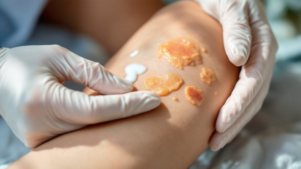
Necrobiosis lipoidica is a rare granulomatous skin disorder strongly linked to diabetes, presenting as yellowish-brown plaques on the shins. First-line treatments include potent topical corticosteroids and intralesional injections, with 40% showing positive results. For resistant cases, photodynamic therapy, biologics, and JAK inhibitors offer alternatives. Vascular medications and surgical interventions may be necessary for severe cases. Proper management requires understanding both dermatological and endocrinological aspects of this complex condition.
Key Takeaways
- Necrobiosis lipoidica is a rare granulomatous skin disorder strongly associated with diabetes, affecting 65% of patients with this condition.
- First-line treatments include potent topical corticosteroids, with alternatives like tacrolimus 0.1% ointment for steroid-resistant cases.
- Photodynamic therapy shows 88% significant improvement rates for resistant cases, typically requiring about 3 weekly sessions.
- Biologics and JAK inhibitors offer powerful alternatives when conventional treatments fail, with TNF-alpha inhibitors showing positive outcomes.
- Surgical intervention may be necessary for severe ulceration, with split-skin grafting demonstrating positive outcomes in multiple studies.
Understanding Necrobiosis Lipoidica and Its Diabetes Connection
While necrobiosis lipoidica represents a rare granulomatous skin disorder first described by Oppenheim in 1929, it’s particularly notable for its strong association with diabetes mellitus. At our Skin and Cancer Institute, we frequently observe this connection, with studies showing 65% of necrobiosis lipoidica patients have diabetes, while many others demonstrate abnormal glucose tolerance.
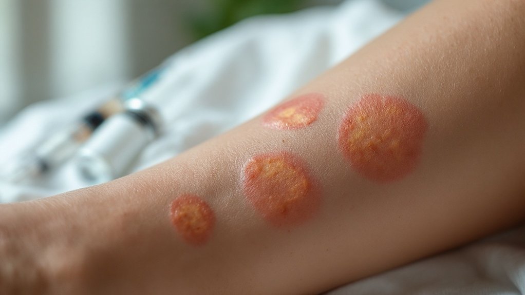
The pathophysiology overview reveals three key components: vascular damage, immune-mediated inflammation, and collagen destruction. Regarding patient demographics, we see this condition mainly affects females at three times the rate of males, with average onset around age 30. Recent multicenter research has demonstrated a more pronounced female to male ratio of approximately 5:1. The disorder typically presents as yellowish-brown plaques with red borders on the shins, progressing to atrophic, telangiectatic patches. In 15% of cases, necrobiosis lipoidica actually precedes diabetes diagnosis, making it an important early indicator.
First-Line Topical and Injectable Treatment Options
We utilize potent topical corticosteroids as our primary first-line treatment for early necrobiosis lipoidica lesions, with intralesional injections reserved for active borders that require more targeted therapy. However, these treatments can worsen skin atrophy, a significant concern for patients with already compromised skin integrity. For patients who don’t respond to steroids or have contraindications, tacrolimus 0.1% offers an effective alternative that prevents ulceration while maintaining disease stability. Cryotherapy can be implemented alongside these approaches, though with more variable results and less robust evidence supporting its effectiveness compared to steroid-based interventions.
Steroid Therapy Benefits
Steroid therapy represents the cornerstone of first-line treatment for necrobiosis lipoidica, offering both topical and injectable delivery options with documented efficacy. Our clinical experience confirms that high-potency formulations like mometasone furoate 0.1% and clobetasol propionate demonstrate superior penetration through sclerotic lesions, achieving positive results in approximately 40% of cases.
For ideal steroid efficacy, we often recommend combination approaches—sequential application of topical steroids followed by intralesional injections for resistant areas. This methodology maximizes therapeutic benefit while supporting patient compliance through visible inflammation reduction. One compelling case involved a patient with a 10-year history of NL who experienced significant temporary improvement with intralesional triamcinolone injections.
We’re particularly attentive to our diabetic patients’ needs, as steroid therapy requires careful monitoring due to potential insulin resistance and blood glucose elevation concerns. For these individuals, we may modify treatment protocols to balance effective lesion management with metabolic stability.
Tacrolimus Alternative Approach
When traditional steroid therapies prove insufficient or contraindicated, tacrolimus 0.1% ointment emerges as our preferred alternative first-line treatment for necrobiosis lipoidica, particularly for patients with early-stage, non-ulcerated lesions.
Our clinical experience mirrors systematic review findings showing efficacy in approximately 65% of cases. We typically recommend twice-daily tacrolimus application for 8 weeks, followed by once-daily maintenance for another 8 weeks. A 67-year-old diabetic woman with atypical NL presentations showed significant improvement after following this exact treatment protocol. For enhanced results, we often implement a dual treatment approach, combining tacrolimus with topical corticosteroids.
This alternative approach proves most beneficial for patients with recent disease onset, often preventing progression to ulceration. At the Skin and Cancer Institute, we carefully assess each patient’s lesion characteristics and disease duration to determine if tacrolimus therapy represents your best treatment pathway.
Cryotherapy Application Methods
Cryotherapy stands as one of our three primary first-line interventions for necrobiosis lipoidica, offering targeted tissue destruction that interrupts the inflammatory cascade responsible for disease progression. While effective for many patients, we recommend implementing specific cryotherapy techniques based on lesion maturity and location.
Our standard application protocols involve liquid nitrogen delivered via spray or contact methods, typically applied in 15-20 second freeze-thaw cycles. For thicker, more established plaques, we’ll often employ multiple freeze-thaw cycles to enhance penetration depth. Peripheral lesions generally respond best to this approach.
At the Skin and Cancer Institute, we customize cryotherapy intensity based on your individual presentation. We’ll carefully monitor your response, adjusting subsequent treatments to balance efficacy with minimal scarring risk. However, patients with diabetes should be cautious as cryotherapy may worsen vascular changes that contribute to NL’s pathogenesis.
Photodynamic Therapy as an Effective Alternative Approach
Photodynamic therapy (PDT) has emerged as a highly effective alternative approach for treating recalcitrant necrobiosis lipoidica, demonstrating impressive response rates between 39% and 90% across multiple published studies. At our practice, we’ve observed that 88% of patients experience significant improvement with PDT treatment.
The procedure typically involves applying methyl aminolevulinate (MAL) or 5-aminolevulinic acid BF-200 as a photosensitizer under occlusive bandaging for 3 hours, followed by red light activation at 632 nm with 37 J/cm² energy density. Most patients require an average of 3.2 weekly sessions. The treatment is particularly valuable for patients who have diabetes mellitus, as this condition is frequently associated with necrobiosis lipoidica.
We’ve found PDT is well-tolerated without analgesia during light exposure. Long-term results are encouraging, with 24-month follow-ups showing stabilized remission in treated patients. Complete clinical and histological resolution can be achieved after approximately 6 treatment sessions.
Biologics and JAK Inhibitors for Treatment-Resistant Cases
For patients with treatment-resistant necrobiosis lipoidica, our dermatology team has increasingly turned to biologics and JAK inhibitors as powerful therapeutic alternatives. These advanced options have demonstrated remarkable efficacy when conventional therapies fail.
TNF-alpha inhibitors (adalimumab, etanercept, and infliximab) often yield positive outcomes in refractory cases. We’ve also seen promising results with dupilumab, which blocks IL-4/IL-13 signaling pathways, achieving full remission in patients with decade-long disease histories. This approach aligns with research highlighting the need for continuous monitoring and reassessment of treatment effectiveness due to the lack of uniform treatment guidelines.
JAK inhibitors represent another breakthrough. Tofacitinib (JAK1/3 inhibitor) notably softens subcutaneous nodules within one month, while topical ruxolitinib (JAK1/2 inhibitor) offers a non-systemic alternative. Additional biologic therapies in our arsenal include ustekinumab, secukinumab, and tapinarof, expanding treatment options for our patients with this challenging condition.
Vascular Medications and Blood Flow Enhancement Strategies
When addressing the complex vascular pathophysiology of necrobiosis lipoidica, our dermatology team employs various medications and strategies designed to enhance blood flow and improve microcirculation.
Our therapeutic approaches target vascular thrombosis through antiplatelet agents like ticlopidine hydrochloride, which reduces platelet aggregation. For microangiopathy treatment, we utilize pentoxifylline and stanozolol to enhance fibrinolysis, counteracting microvascular occlusion mechanisms common in diabetic skin lesions. Studies indicate that patients with necrobiosis lipoidica show altered cutaneous microcirculation, which substantiates the need for blood flow enhancement therapies.
We’ve found that vasodilatory compounds such as inositol niacinate and nicofuranose provide vascular smooth muscle relaxation, addressing the reduced perfusion in affected areas. For ulcerative complications, hyperbaric oxygen therapy increases tissue oxygenation to support cellular repair.
While individual responses vary, we often combine these vascular strategies for synergistic effects in treatment-resistant cases, creating thorough protocols that optimize blood flow to damaged skin.
Surgical Management and Handling Complications

Surgical intervention becomes necessary in cases where necrobiosis lipoidica progresses to severe ulceration or remains resistant to conservative therapies. At our practice, we employ thorough surgical excision down to the fascia level followed by split-skin grafting for severe cases. This approach has demonstrated positive outcomes in multiple studies, including research from Stanford University Medical Center showing no recurrence in seven cases. Tissue-engineered dermal skin grafting has shown promising results as an alternative when conventional grafting isn’t feasible.
Ulcer management requires specialized wound care following diabetic ulcer principles. We’re vigilant about complications, as chronic ulcerative lesions can develop squamous cell carcinoma. Post-surgical prognosis includes potential recurrence, with appearance normalization requiring extended timeframes. We emphasize prevention strategies and glucose monitoring throughout treatment. For complex cases requiring surgical excision or ulcer management, our dermatology team provides extensive, personalized care.
Frequently Asked Questions
How Long Will Necrobiosis Lipoidica Last if Left Untreated?
Without treatment, necrobiosis lipoidica typically persists indefinitely—often for life. We recommend exploring treatment options and specialized skin care rather than leaving this chronic condition untreated, as spontaneous resolution occurs in less than 20% of cases.
Can Diet Modifications Improve Necrobiosis Lipoidica Symptoms?
We’ve found limited evidence that dietary changes affect necrobiosis lipoidica. While antioxidants and low-fat diets have been attempted, documented nutrient impact remains insufficient to recommend specific modifications for symptom improvement.
Are Children With Diabetes at Risk for Necrobiosis Lipoidica?
Yes, children with childhood diabetes are at risk for necrobiosis lipoidica. We’ve observed this skin complication typically developing after several years of diabetes, often associated with other microvascular complications in our pediatric patients.
Does Necrobiosis Lipoidica Increase Infection Risk During Normal Activities?
Yes, necrobiosis lipoidica greatly increases infection risk during normal activities. The thin, atrophic lesion centers can easily break open from routine friction, requiring vigilant infection prevention strategies during daily activities we all perform.
Can Necrobiosis Lipoidica Recur After Successful Treatment?
Yes, necrobiosis lipoidica can recur after successful treatment. We’ve observed recurrence factors including diabetes status, female gender, and pretibial location. Multiple treatment options are often needed for recurring cases.
Conclusion
We’ve found that successful management of necrobiosis lipoidica requires a multifaceted approach. We’ll integrate topical corticosteroids, intralesional injections, phototherapy, and systemic agents as indicated by lesion characteristics and disease progression. When ulcerations develop, we implement specialized wound care protocols. Ongoing glucose control remains essential, as we’ve observed correlation between hemoglobin A1c levels and treatment response. Regular monitoring every 3-6 months allows for timely therapeutic adjustments.